Features of the TS at the fabric level, thread level, and fiber level…
At the fabric level:
The image does not go through the entire cloth, but only resides on the surface. It is not a result of any pigment, dye, paint or stain, but from dehydration/oxydation of the fabric.
No pigments, paints, dyes or stains have been found on the fibrils. X-ray, fluorescence and microchemistry on the fibrils preclude the possibility of paint being used as a method for creating the image.The scientific consensus is that the image was produced by something which resulted in oxidation, dehydration and conjugation of the polysaccharide structure of the microfibrils of the linen itself.
https://www.shroud.com/78conclu.htm
The color only resides on the external surface of the
TS.The superficial color is not due to any pigment
since no pigment particles can be seen either mac-
roscopically or microscopically nor are there any
external substances or evidence of media scorching
in image areas. The color is only due to a chemical
reaction (dehydration and oxidation).
https://www.academia.edu/4294684/Micros … rficiality
There is no evidence of any cementation on the cloth.
There is no cementation signs among the image fibers
https://shroud.com/pdfs/doclist.pdf
There is no dry power on the cloth.
There is no observed microscopic, chemical, or spectroscopic evidence for the presence of
any dry powder responsible for the body image on the TS
https://shroud.com/pdfs/doclist.pdf
https://www.academia.edu/4294684/Micros … rficiality
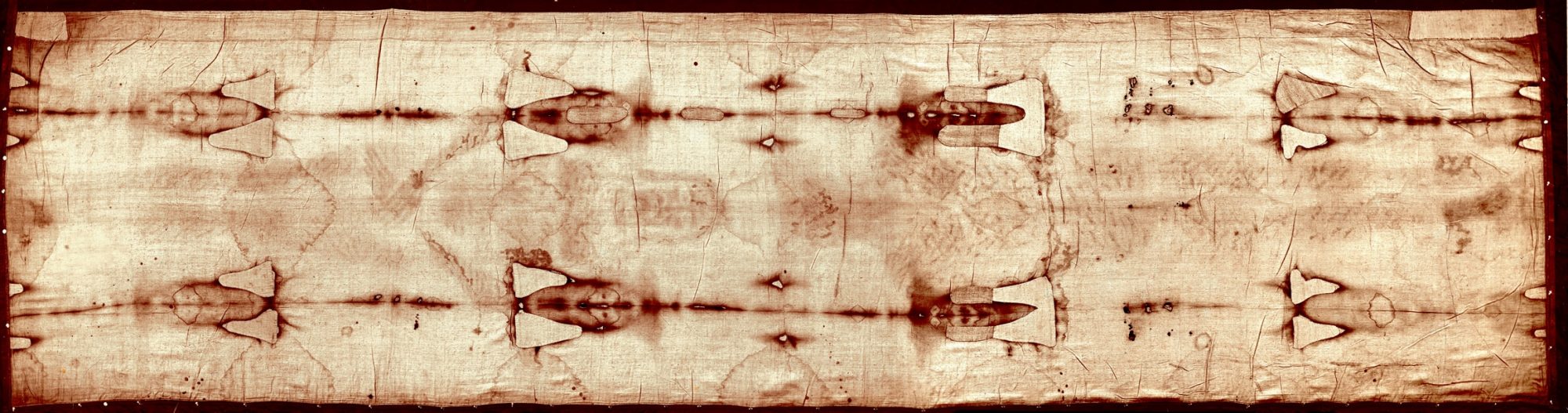
There is little variation between the color density and distribution on the dorsal and ventral sides of the body.
The image of the dorsal side of the body shows the same color density and distribution as the
ventral, and it does not penetrate the cloth any more deeply than the image of the ventral side
of the body.
https://www.shroud.com/pdfs/rogers5faqs.pdf
The image of the dorsal side of the body shows
nearly the same color density and distribution as
the ventral, but the Face image shows a higher color
density.
https://www.academia.edu/4294684/Micros … rficiality
The second major aspect of the uniformity feature, relates to the fact that STURP scientists have proven that both the Shroud’s frontal and dorsal body images have approximately the same maximum optical densities (or colour darkness); we shall call this the density uniformity aspect. To be more specific, both the frontal and dorsal body images are very faint; each having typical reflected optical densities of less than 0.1 in the visible range. As such, this aspect seems to pose an initial problem for some of “ordinary” naturalistic theories such as ones involving the presence of a dead corpse and/or human-size statue laying in a supine position while wrapped in the Shroud. The reason this aspect poses a problem to such mechanism theories is because, in the words of Shroud expert Serge Mouraviev, “the dorsal side of the body must have been gravitationally pressed to the cloth by its weight with an estimated average pressure of 26.8 g/cm2 as against only 0.35 g/cm2 for the pressure of the cloth on the contact areas of the frontal side of the body”. Common sense then, would indicate that one might expect the dorsal side image to be visibly darker than the frontal image if such image-forming hypotheses were true, yet this is not what we find with the Shroud of Turin’s body images.
https://realseekerministries.files.word … apter.docx
At the thread level:
Only the top one to three fibers of a thread are discolored.
The color only resides in the most external (two or
maximum three) fibers of the threads.
https://www.academia.edu/4294684/Micros … rficiality
Colored fibers are alongside non-colored fibers.
Some noncolored fibers in image areas can be
found adjacent to colored TS image fibers on a
given thread of the image areas.
https://www.academia.edu/4294684/Micros … rficiality
At the fiber level:
A discolored fiber is uniformly colored.
If a fiber is colored, it is uniformly colored around its cylindrical surface (Adler 1996, 1999)
https://shroud.com/pdfs/doclist.pdf
Only the exterior of a fiber is discolored. The interior of a fiber is not affected.
The image only resides in the external surface prob-
ably corresponding to the “primary cell wall”
composed of polysaccharides of lower activation
energy than the cellulose.The cellulose of the linen fiber residing in the “sec-
ondary cell wall” is not colored and the medullas
of the 10– 20- m-diameter fibers in image areas
also appear colorless.
https://www.academia.edu/4294684/Micros … rficiality
Any image-formation mechanism that would result in color formation inside the linen
fibers must be rejected. Some “theories” that have been mentioned that would cause coloration
inside fibers are penetrating radiation, high temperature scorching (hot statue, painting with a
torch, etc.), and catalyzed dehydration of the cellulose. Image fibers are colored only on their
surfaces.Image color can be chemically reduced with diimide, leaving colorless cellulose fibers. All
image color resides on the outer surfaces of the fibers.The medullas of colored image fibers are not colored: The cellulose was not involved in
color production.
https://www.shroud.com/pdfs/rogers5faqs.pdf
https://debatingchristianity.com/forum/viewtopic.php?p=1123427#p1123427